Trained Neural Networks (U-Net) and Instructional Videos
Instructional videos
These videos are indented to allow interested labs to set up and run deep learning tools for analysis of phenotypes in developmental biology. The Fiji-plugin that was used to train and deploy networks is described inFalk et al., Nature Methods, 2019.
If you use any of the pre-trained weight files in your own work, please cite: Naert et al., Development, 2021, https://doi.org/10.1242/dev.199664
Pre-trained weight files
Here, we provide pre-trained weight files for various U-Nets. These were trained to recognize, segment or classify various structures in 2D or 3D volume stacks.
To run each network, please download the zip archive, which contains:
a model file (modeldef.h5)
the weights file of the trained model (caffemodel.h5)
a sample input file
the corresponding output file
a short description of the network (readme file)
2D-BrainNet
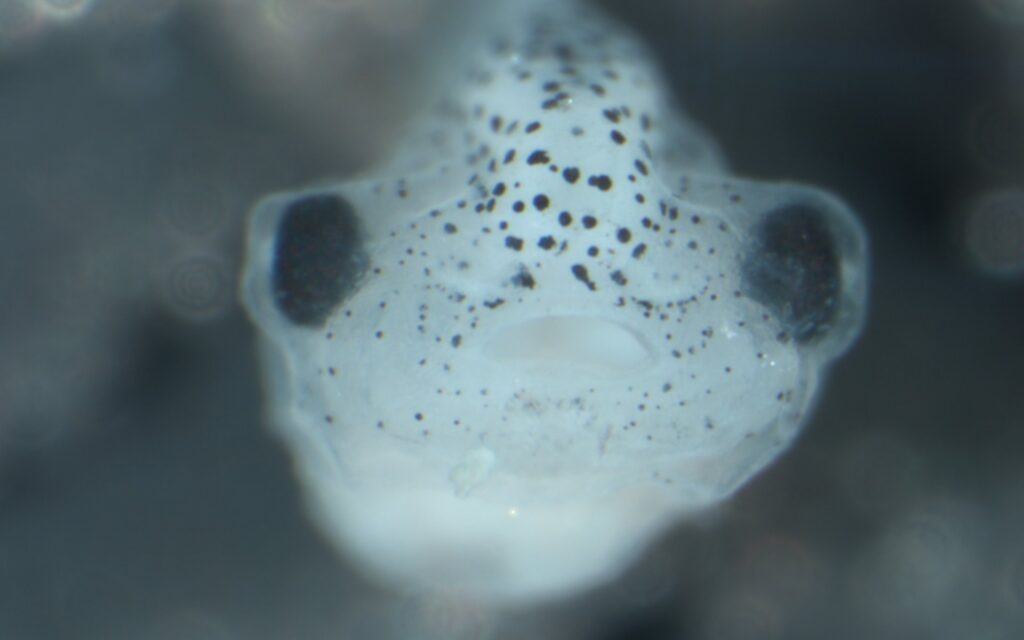
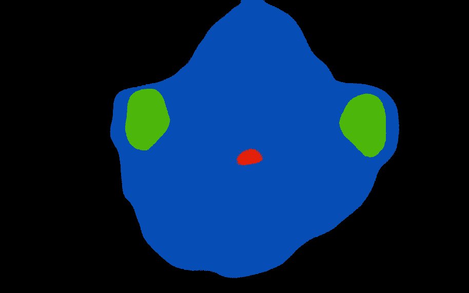
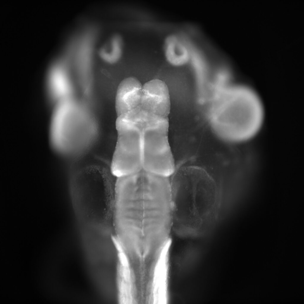
2D-BrainNet is a U-Net model for multiclass segmentation of different brain regions in both hemispheres, of Beta-Tubulin-stained X. tropicalis embryos. This model was validated on X. tropicalis embryos.
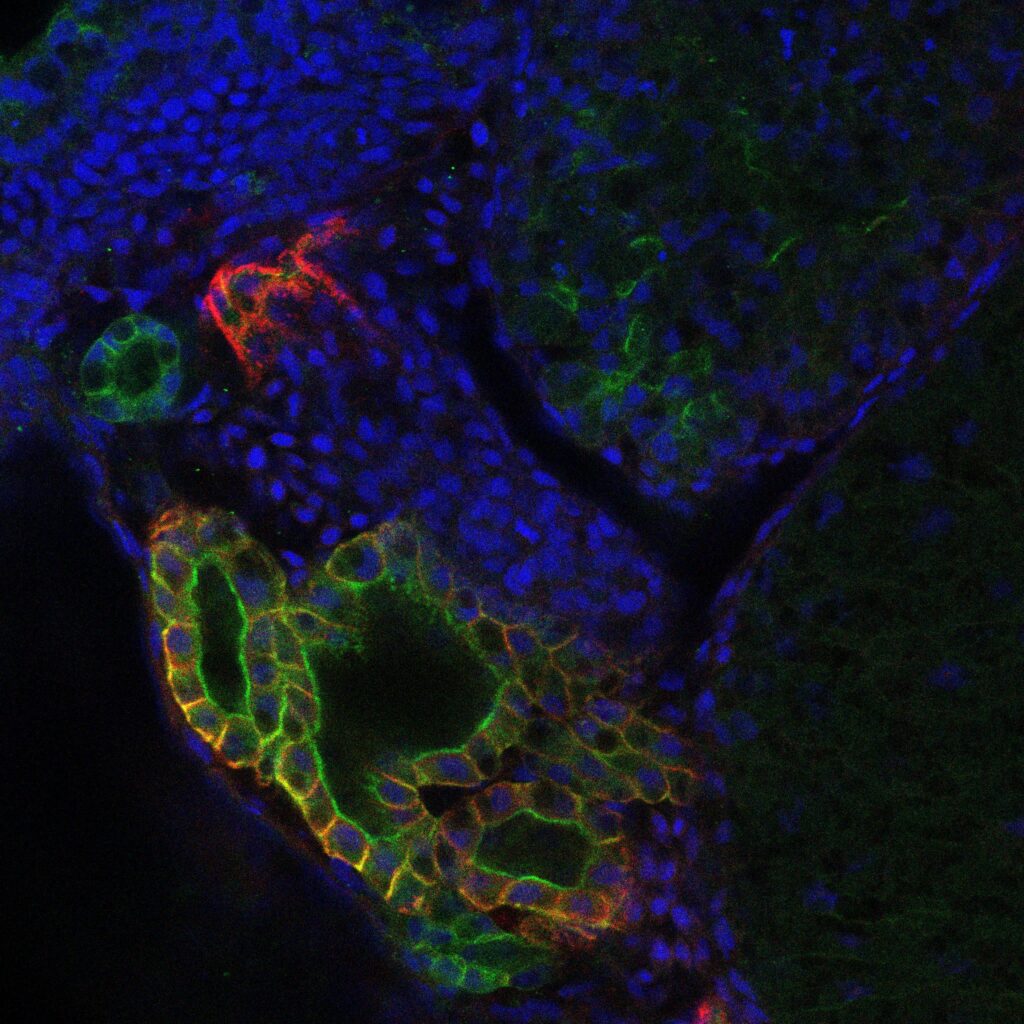
2D-CystNet is a U-Net model for classifying X. tropicalis pronephros into either the class “normal” or the class “cystic”. Input images should contain only the pronephros, surrounded by a white background. This model has been deployed on masks generated by 2D-NephroNet and has been validated on three distinct stages of X. tropicalis development (st. 33, st. 38 and st. 41)
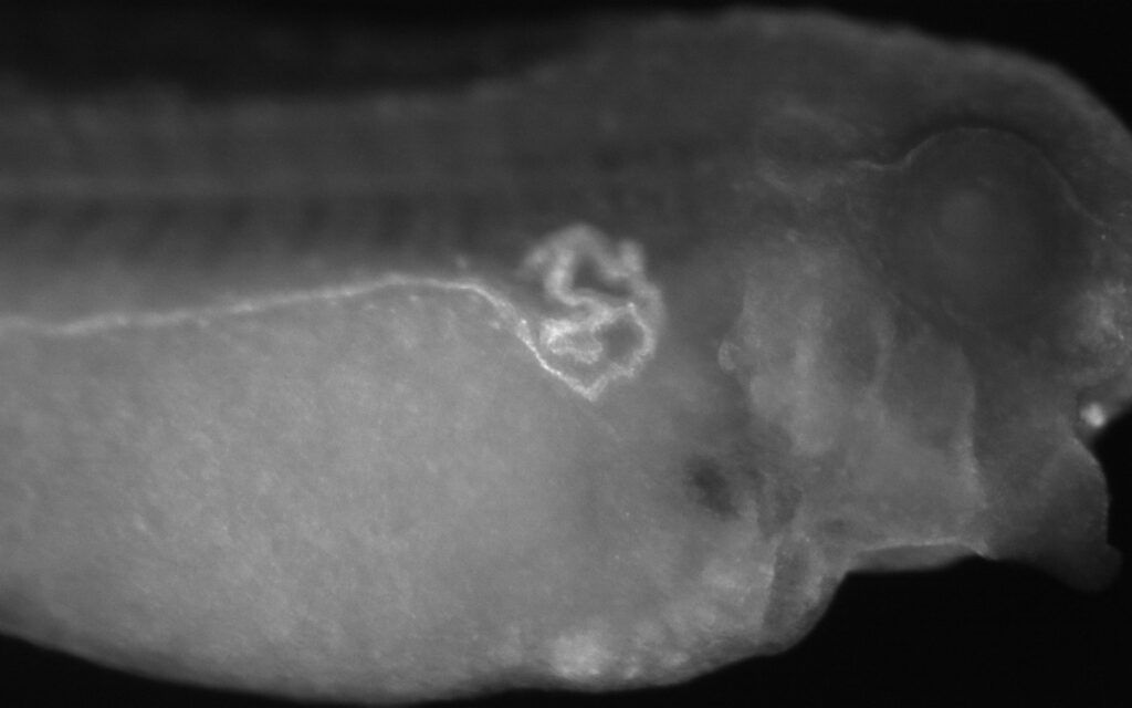
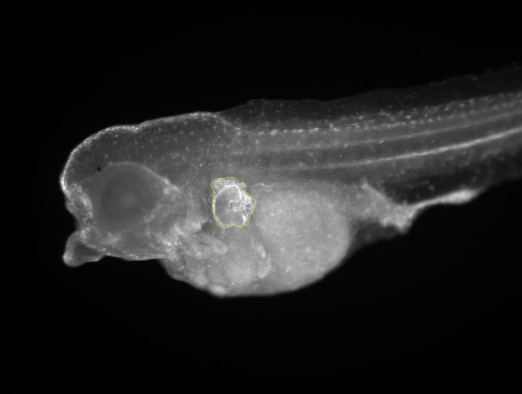
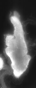
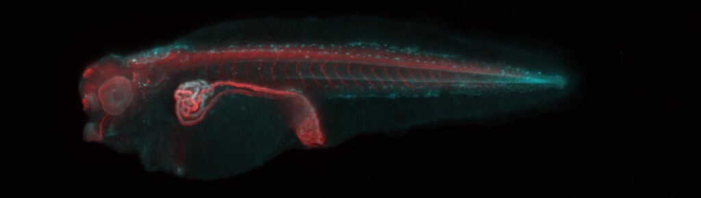
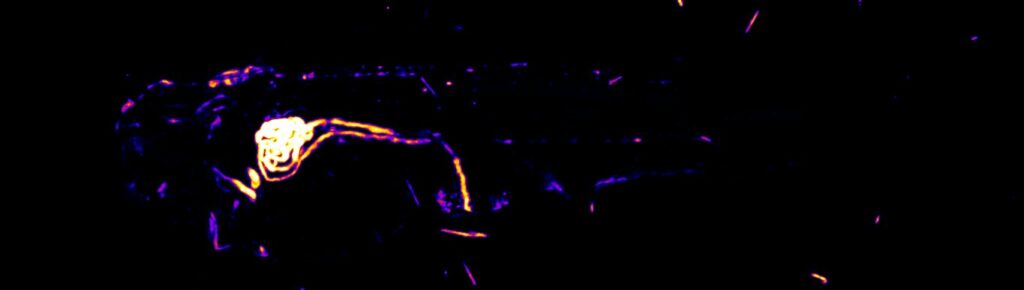
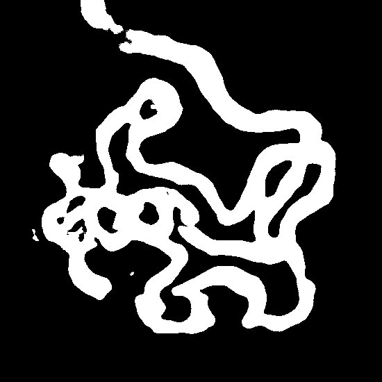
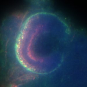
2D-NephroNet is a U-Net model for segmentation of pronephros from LEL-Lectin stained X. tropicalis embryos. This model has been validated for hypoplastic, normal and cystic kidneys in stage 38 X. tropicalis embryos.
2D-NephroNet is a U-Net model for segmentation of pronephros from LEL-Lectin stained X. tropicalis embryos. This model has been validated for hypoplastic, normal and cystic kidneys across three stages of development in X. tropicalis embryos (st. 33, st. 38 and st. 41).
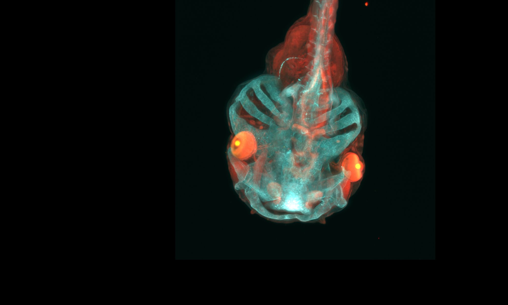
3D-BrainNet is a U-Net model for 3D reconstruction of the brain from a Beta-Tubulin stained stage 45 X. tropicalis embryo. A MesoSPIM recording was sparsely annotated (10% of slices) for training, allowing three-dimensional reconstruction. This model was not validated beyond this single sample, but weights can serve as a baseline for further finetuning approaches.
3D-NephroNet is an U-Net model for 3D reconstruction of the pronephros from stage 38 LEL-lectin-stained X. tropicalis embryos imaged by MesoSPIM light-sheet microscopy. This model was validated in unseen MesoSPIM recordings.
3D-CystNet is a U-Net model for 3D reconstruction of both the kidney epithelium as well as possible cysts from confocal imaging stacks. This model was validated on a stack from a normal and a cystic kidney.
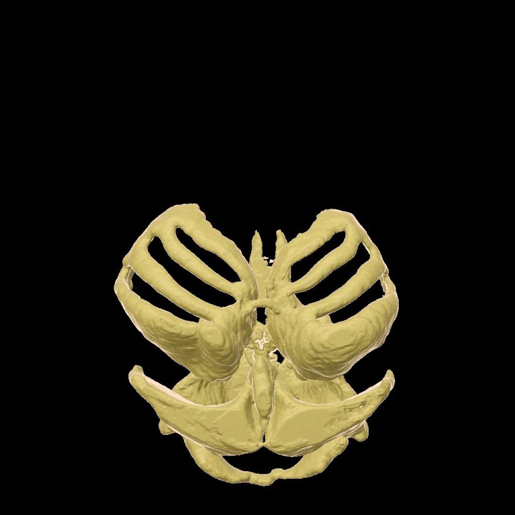
CranioNet is a U-Net model for 3D reconstruction of the craniofacial cartilage from col2a1-stained X. tropicalis embryos imaged by MesoSPIM light-sheet microscopy. This model was validated in unseen MesoSPIM recordings.
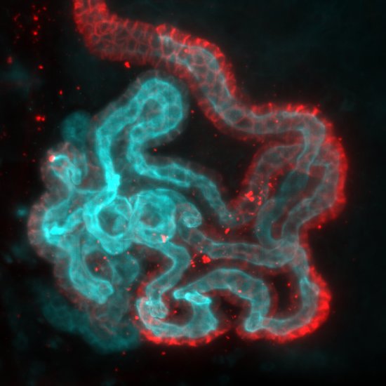
DCT-Net is a U-Net model for 3D reconstruction of the distal convoluted tubules (DCT) in CLARITY-cleared kidneys from a NCCcre/+:TdTomato-redTg/+ male mouse.
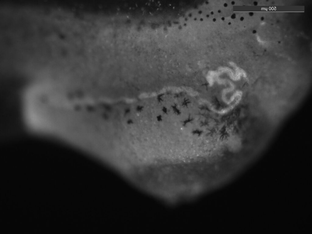
DiameterNet is a U-Net model for segmenting the kidney tubule from lectin/A5-stained embryos, imaged using high-resolution MesoSPIM. Input is a maximum intensity projection of the pronephros.
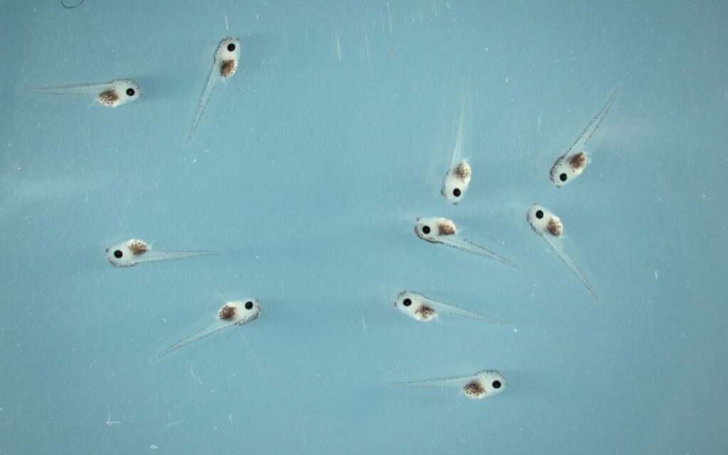
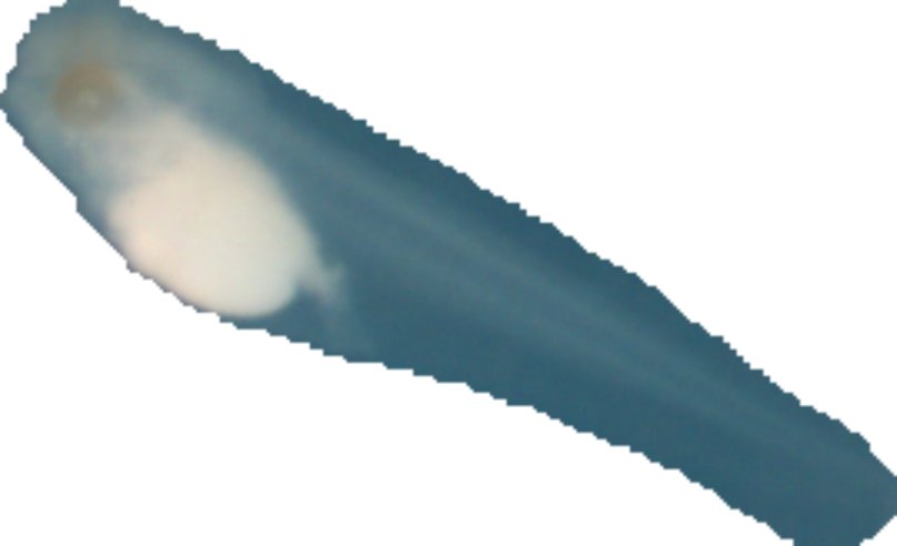
EmbryoNet is a U-Net model for segmentation of single embryos from low magnification stereomicroscopy images, containing multiple embryos in a single field-of-view.
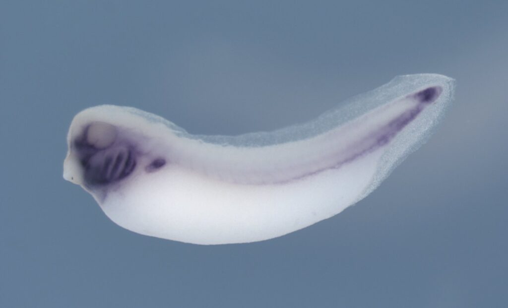
EmbryoNet-ISH is a U-Net model for segmentation of in situ hybridization-stained (ISH) X. tropicalis embryos imaged on agar plates by stereomicroscopy. Model is naïve to the employed ISH probe and to the developmental stage. Model has been validated across X. tropicalis developmental stages ranging from stage 10 to stage 40.
EyeNet is an U-Net model for eye segmentation in X. tropicalis embryos. Input images should contain a single embryo, surrounded by a white background. This model has been deployed on masks generated by EmbryoNet and is naïve to potential loss of pigment due to tyrosinase genome editing.
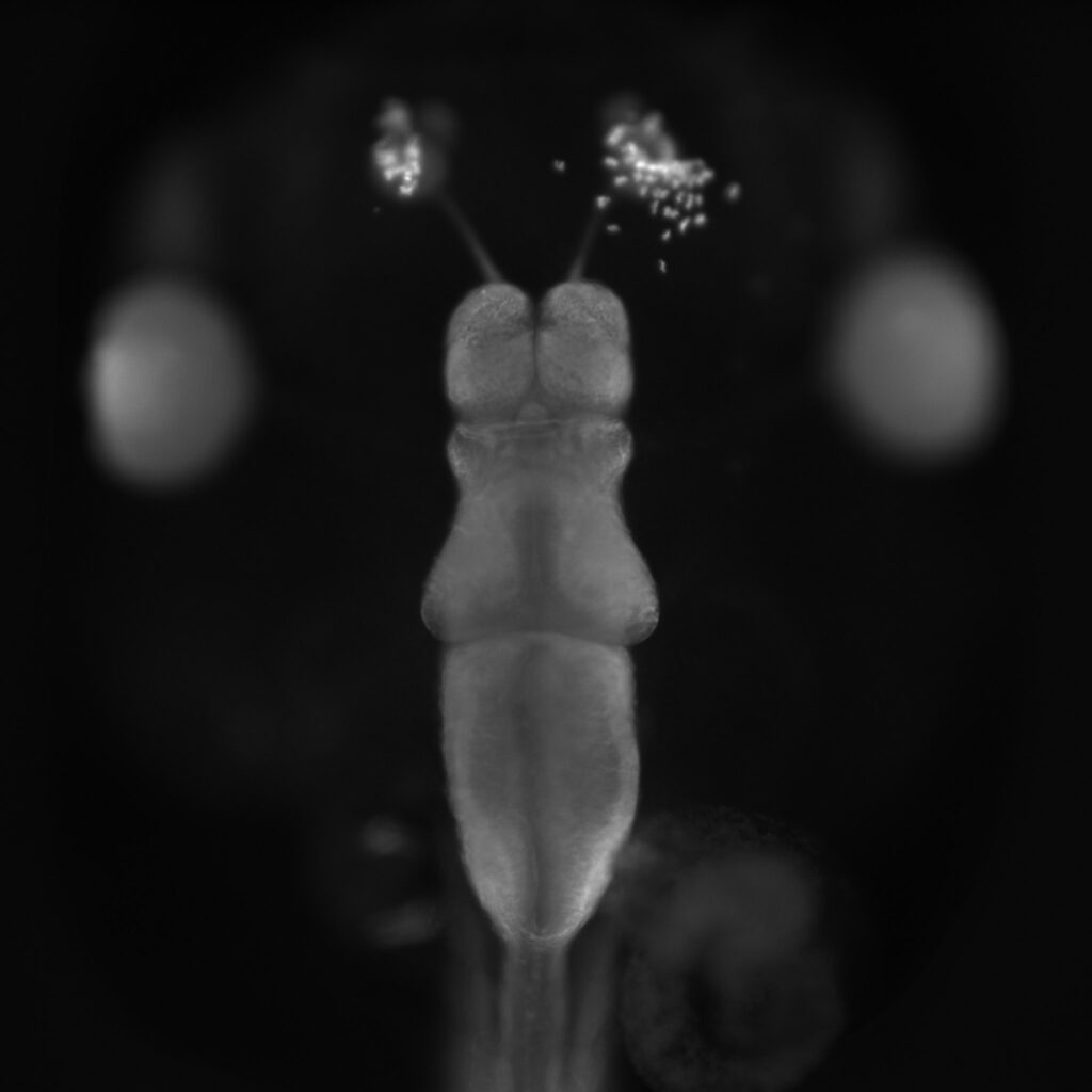
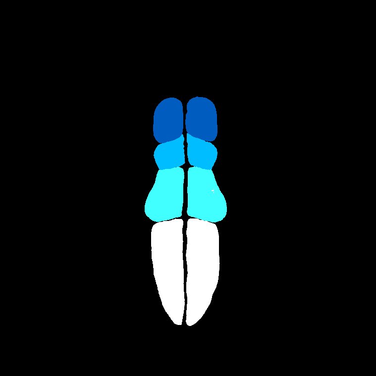
FaceNet is a U-Net model for multiclass segmentation of different orofacial regions in X. tropicalis embryos imaged by bright-field stereomicroscopy. The model is naïve to perturbations occurring in response to chemical treatments with the BMS-453 inhibitor.
OrganNet is an U-Net model for organ segmentation in X. tropicalis embryos. Input images should contain a single embryo, surrounded by a white background. This model has been deployed on masks generated by EmbryoNet and is naïve to potential loss of pigment due to hydrogen peroxide bleaching.
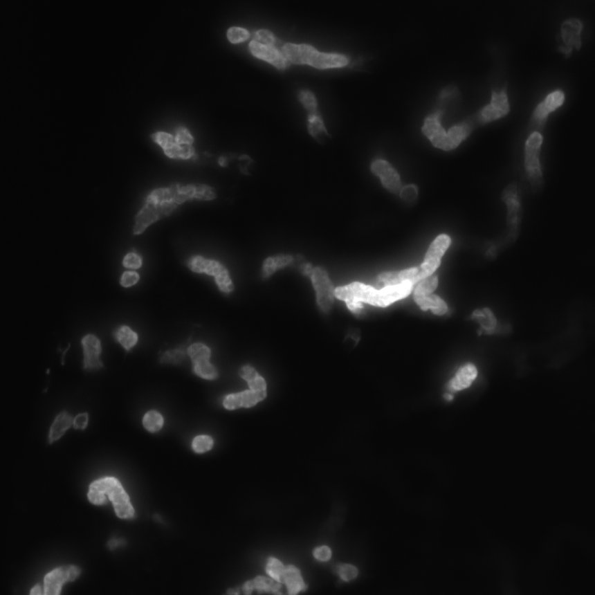
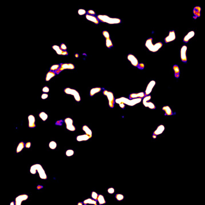
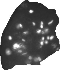
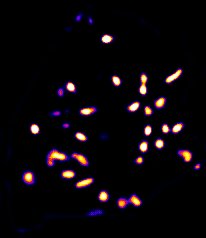
ProliNet is a U-Net model for segmentation of cells in the S-phase of cell division, as identified by positive staining for the marker phospho-histon H3, in X. tropicalis telencephalon. Input images should contain only the pronephros, surrounded by a white background. This model has been validated on masks generated by TelenNet.
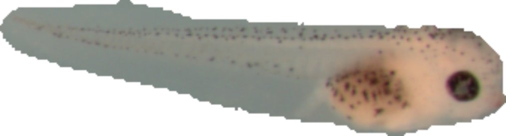
TelenNet (Beta-Tubulin) is a U-Net model for segmentation of telencephalon from beta-tubulin stained X. tropicalis embryos. This model has been validated for normal brains and for brains affected by dyrk1a loss-of-function.
TelenNet (PCNA) is a U-Net model for segmentation of telencephalon from proliferating cell nuclear antigen (PCNA) stained X. tropicalis embryos. This model has been validated for normal brains and for brains affected by dyrk1a inhibition via Harmine.
TubuleNet is a dense U-Net model for segmentation of pronephric tubules from LEL-Lectin stained Xenopus embryos. This model was validated for hypoplastic and normal kidneys in stage 38 X. laevis embryos.
MabNet is a U-Net model for segmentation of retina and lens from Atp1a1, lectins peanut agglutinin (PNA) and wheat germ agglutinin (WGA) stained X. tropicalis embryos.